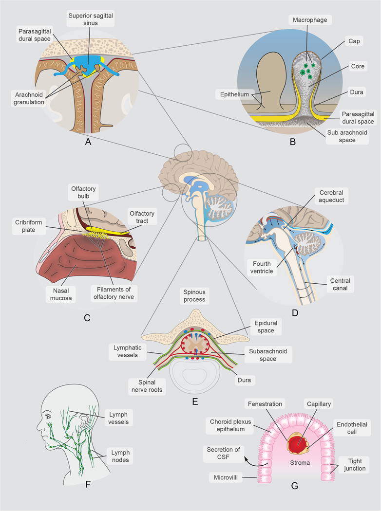
Fig. 1. Anatomical Points of Interest in CSF Circulation and Glymphatic Flow.
Important points of interest labeled:
a) Cross section of the superior sagittal sinus region, showing the superior sagittal sinus, parasagittal dural space (PDS), and arachnoid granulations
b) Anatomy of an arachnoid granulation, where subarachnoid flow of CSF reabsorbs into dural venous sinuses
c) Cranial nerves, showing location of CSF flow around cranial nerves (i.e. cribiform plate, CNI)
d) Cerebral aqueduct and 4th ventricle, detailing the path of CSF flow from the third ventricle to the central canal and subarachnoid space
e) Axial cross-sectional anatomy of the spinal canal with the relationship of the lymphatic vessels to the thecal sac, epidural space and spinal nerve roots.
f) Cervical lymphatic flow, detailing the lymph vessels and nodes.
g) Choroid Plexus, predominantly located in the lateral ventricles, where CSF is produced and secreted in the ventricular system.

