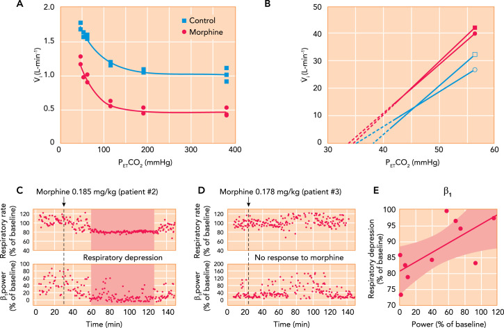
FIGURE 5.
Contributions of chemodrive and “awake drive” to minute ventilation and opioid effects
A: hypoxic ventilatory response curves in a chloralose-urethane anesthetized cat during control (solid square) and after administration of 0.15 mg/kg IV morphine. Morphine decreased minute ventilation during hyperoxia but did not change the increase in minute ventilation with hypoxia (see Ref. 12). B: mean ventilatory response to increasing inspiratory carbon dioxide concentrations obtained in 12 male (square) and 12 female (circle) human volunteers during control (filled) and after 0.1 mg/kg IV morphine, followed by 0.03 mg · kg–1 · h–1 (open). The continuous lines are the linear regression lines through the data points, and broken lines are extrapolated to the apneic threshold. In men and women, morphine decreased minute ventilation differently: in men by increasing the apneic threshold but in women by decreasing carbon dioxide sensitivity (see Ref. 30). C–E: association between respiratory depression from analgesic doses of morphine and loss of “awake drive” per electroencephalogram in pediatric patients. C: after 0.185 mg/kg morphine, patient 2 presented substantial respiratory rate depression associated with a decrease in β1 power. D: in patient 3, a similar dose (0.178 mg/kg) did not reduce β1 power or cause notable respiratory depression. E: in 10 patients, the severity of respiratory rate depression correlated with the intensity of the reduction in β1 power (R = 0.715, P = 0.02) (105).

