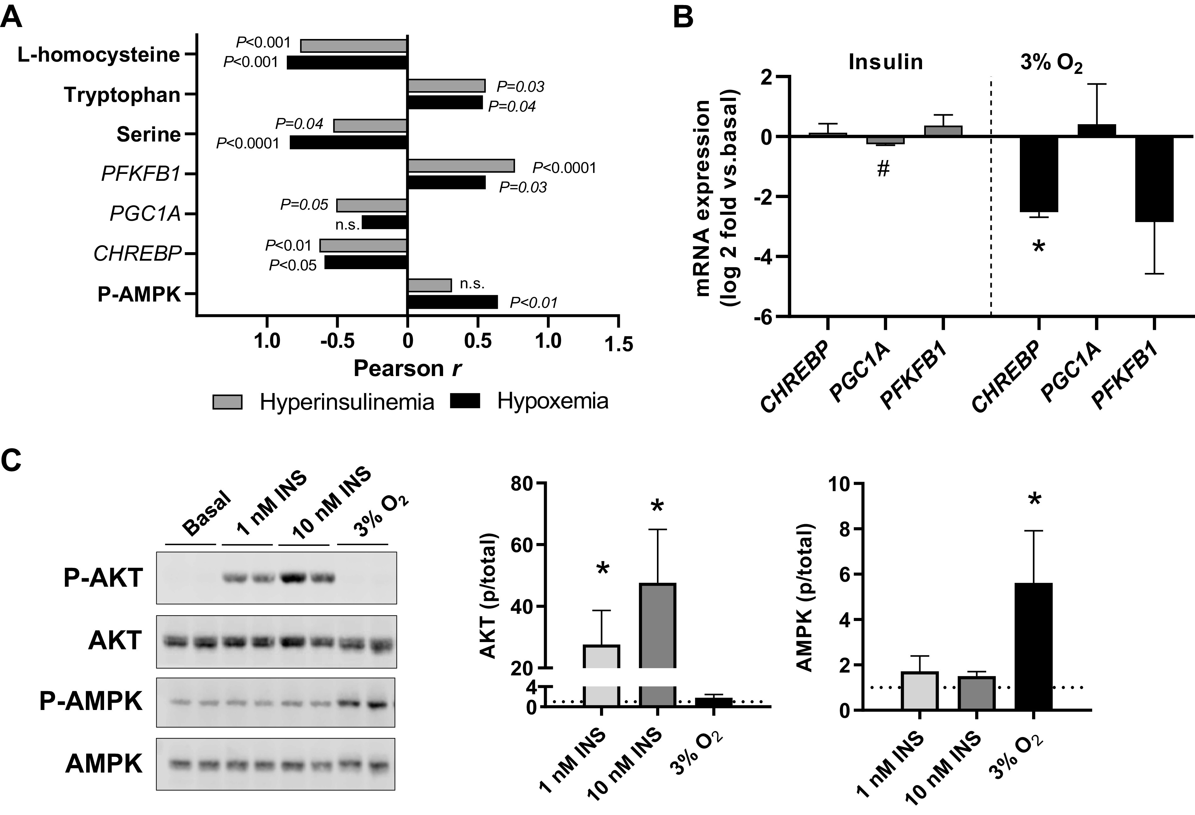
Fig. 7.

Role of hyperinsulinemia and hypoxemia on metabolic and signaling effects in the hyperinsulinemic (INS) fetal liver. A: the association between key metabolites, genes, and phosphorylation of AMP-activated protein kinase (p-AMPK) with hyperinsulinemia (gray bars) or degree of hypoxemia (black bars) represented by multiplying fetal oxygen concentrations by −1. Correlation coefficients are shown. B: isolated primary fetal hepatocytes were treated for 24 h with 100 nM insulin (at ambient air conditions) or 3% oxygen (3% O2). RNA was isolated and gene expression measured. Results are expressed as Log2 fold change relative to a basal treatment (represented by 0 in graphs) in ambient air. Means and SE shown (n = 2 primary cell preparations). #P < 0.10 and *P < 0.05 using 1-sided t test relative to basal treatment. C: isolated primary fetal hepatocytes were studied under basal conditions in ambient air (Basal) and compared with hepatocytes treated with insulin at 1 and 10 nM doses for 30 min or hypoxia with 3% O2 for 4 h. Samples were analyzed by Western blotting for phosphorylated (p-Akt) and total Akt (S473) and AMPK (T172) protein expression. Means and SE shown (n = 2 primary cell preparations). *P < 0.05 relative to basal treatment, indicated with dotted line equal to 1.0. CHREBP, carbohydrate-responsive element-binding protein; PGC1A, peroxisome proliferator-activated receptor-γ coactivator-1α; PFKFB1, 6-phosphofructo-2-kinase/fructose-2,6-biphosphatase 1.
