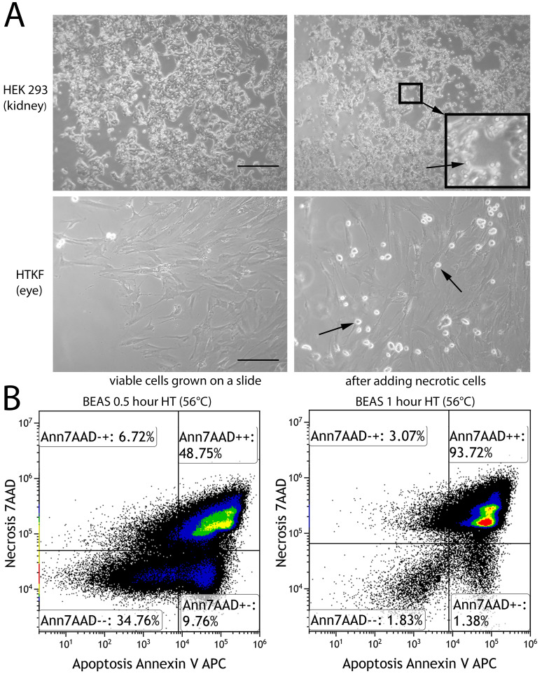
Fig 1. Induction of non-professional phagocytosis by hyperthermia.
(A) Microscopic images (100x magnification) of kidney cells and human Tenon’s capsule fibroblasts grown in coherent cell layers prior to and after adding necrotic, homotypic cells. Arrows are pointing at round shaped necrotic cells. Scale bars are 100μm. (B) Flow cytometric analysis of human bronchial epithelium cells (BEAS) after hyperthermia treatment for half an hour and one hour duration. Annexin V-APC and 7-amino-actinomycin D were used to detect apoptotic and necrotic cells, respectively.

