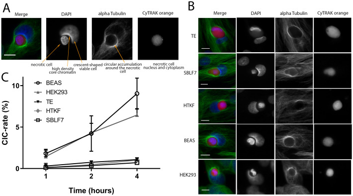
Fig 2. Non-professional phagocytosis in mesenchymal and epithelial cells.
(A) Criteria for CIC evaluation are examplarily shown for a human bronchial epithelial cell (BEAS). Nuclei staining is performed with DAPI (blue), cytoskeleton with alpha-tubulin (green) and necrotic cell with CyTRAK orange (red). (B) CIC formation in the studied cell lines in confluently grown cell layers. Scale bars indicate 10 μm (C) Non-professional phagocytosis rates of the five studied cell lines are calculated as a ratio of observed cell-in-cell structures to all detected viable cells. All experiments were repeated at least three times and not less than 1000 viable cells were counted.

