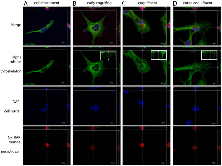
Fig 3. Different phases of non-professional phagocytosis.
Four different phases of cell-in-cell structure formation. α-tubulin was used for cytoskeleton staining, DAPI for cell nuclei and CyTRAK orange for necrotic cell staining. Using the microscope’s Z-stack imaging sequence enabled a representation in XYZ planes. (A) Cell attachment, (B) early engulfing cell, (C) engulfment and (D) late entire engulfment of the dead (red) cell by the living cell (green). Enlarged sections display the reconstruction of α-tubulin cytoskeleton around the necrotic cell.

