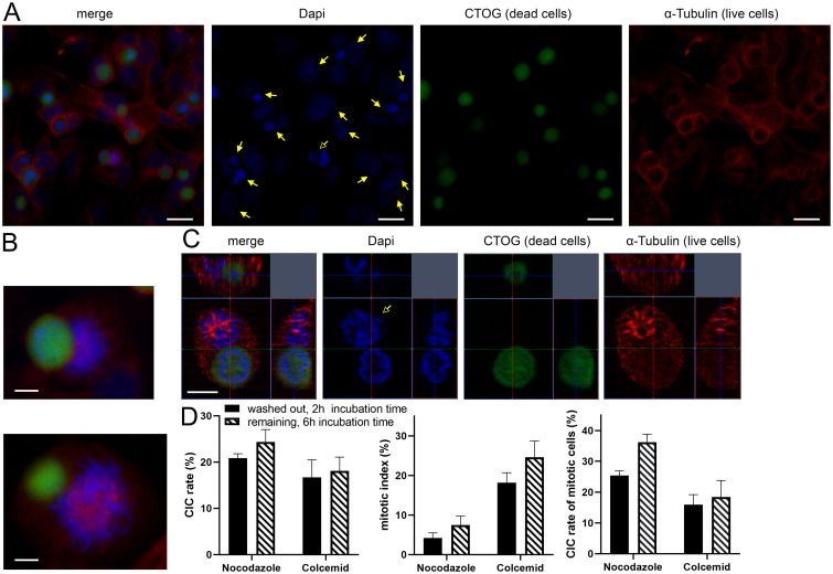
Fig 7. Nocodazole and Colcemid increase mitotic CIC rates in BEAS.
(A) Overview scan of a human lung cell layer treated with Nocodazole for 16 hours. DAPI was used for cell nuclei staining, CTOG for necrotic cells and α-tubulin for cytoskeleton. Filled arrows indicate CIC-structures, unfilled arrows points to mitotic cell involved in non-professional phagocytosis. Scale bars: 10μm. (B) Mitotic cells involved in non-professional phagocytosis. Scale bars: 5μm. (C) Three-dimensional microscopic image of a mitotic cell that has completely absorbed a necrotic cell in its cytoplasm. Scale bars: 5μm. (D) The left bar chart maps CIC-rates of BEAS after treatment either with Nocodazole or Colcemid for 16 hours. Middle: mitotic index of lung cells, which was calculated as the proportion of mitotic cells in all living cells. Right bar chart: Mitotic cells involved in CICs, calculated as proportion of mitotic cells that have incorporated a necrotic cell compared to all mitotic cells.

