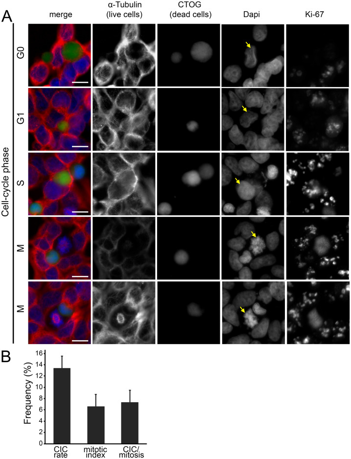
Fig 8. CICs as a function of all cell cycle phases in HEK293 cells.
(A) Fluorescence microscopy images of cell-in-cell structures from various cell-cycle phases. Cell nuclei staining was performed by DAPI, α-tubulin was used for cytoskeletal staining, dead cells were stained by CTOG and Ki-67 served as proliferation marker. Ki-67 was not included in the merged image. Scale bars: 5μm. (B) CIC-rates, mitotic index and mitotic cells involved in CICs of HEK293 cells after treatment with Nocodazole for 16 hours.

