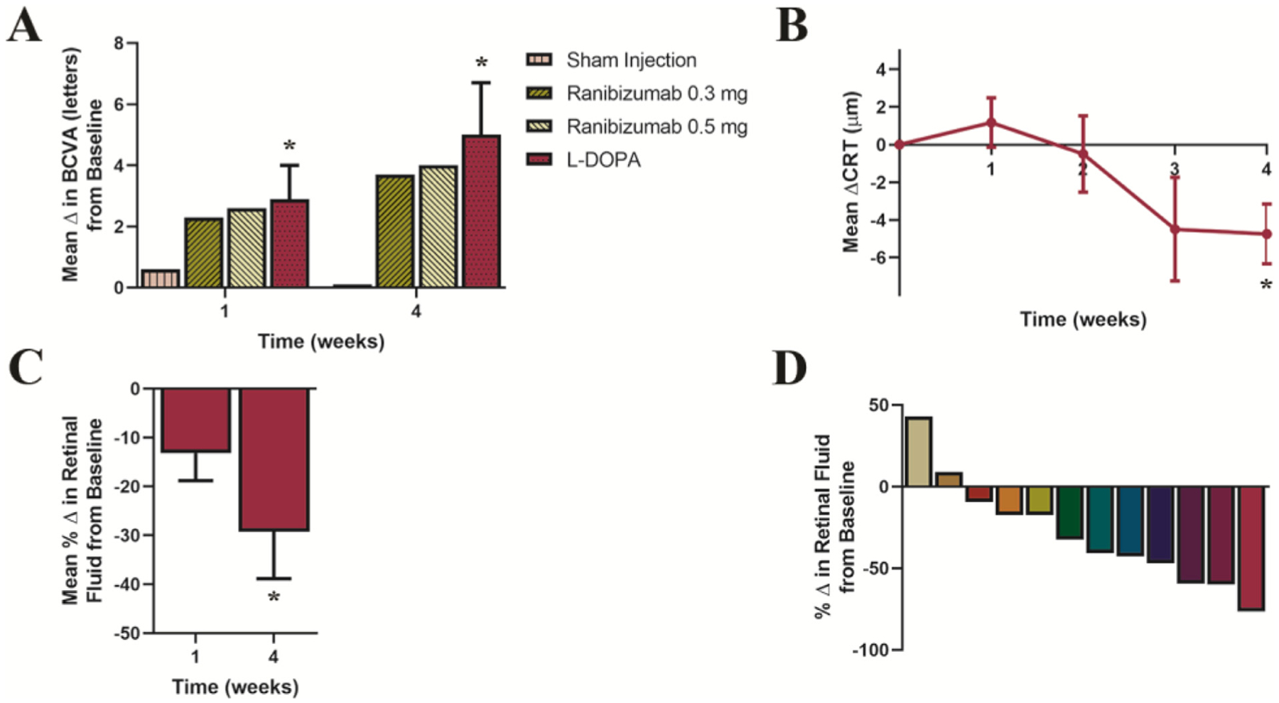
Figure 1.

Changes from baseline in best-corrected visual acuity (BCVA), central retinal thickness (CRT), and retinal fluid in patients naïve to intravitreal anti-vascular endothelial growth factor therapy (Cohort 1). (A) Mean change from baseline in BCVA ± SE during a 1-month period of levodopa treatment and for comparison, the reported changes in visual acuity adapted from a multicenter, 24-month, sham-controlled study in patients receiving intravitreal injections of ranibizumab (0.3 mg or 0.5 mg).28 Starting from a baseline mean of 43.4 letters (20/40), at week 1, BCVA increased by 2.9 letters, P = .03; and at week 4, BCVA increased by 5.0 letters with levodopa treatment, P = .03. (B) Mean change from baseline in CRT ± SE over time, CRT decreased by 4.8 μm, P = .02. Wilcoxon-matched pairs signed rank analyses were used to assess changes in BCVA, and paired-sample t tests were used for changes in CRT. (C) Mean percentage change from baseline in retinal fluid ± SE at 1 and 4 weeks. Mean retinal fluid decreased 13% by week 1, P = .07 and by 29% at week 4 (P = .01; 95% CI, −50.3% to −8.2%). To evaluate percentage change in retinal fluid at weeks 1 and 4, from a theoretical mean of no observed change (retinal fluid = 0.0), one-sample t tests were used. (D) Percent change in retinal fluid at 4 weeks in individual patients. Statistical tests in this figure were 2-sided and adjusted for multiple comparisons with Holm-Bonferroni correction, n = 12.
