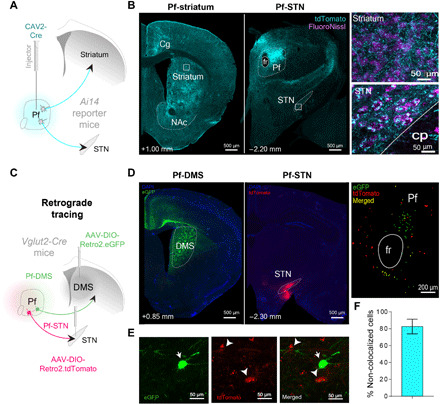
Fig. 1. Separate populations of Pf neurons project to the striatum and STN.

(A) CAV2-Cre was injected into the Pf of Ai14 transgenic mice to visualize afferent and efferent projections. Injection of high-titer CAV2-Cre allows visualization of both input and output connectivity by uptake both in the retrograde and anterograde directions. Neurons in Ai14 mice fluoresce (tdTomato, cyan) when infected with a virus with Cre recombinase. (B) Pf terminal labeling in the striatum and STN. Insets show high-magnification view of Pf terminal labeling. (C) Cre-dependent retrograde viruses were injected into both the DMS and STN of Vglut2 mice (n = 3) to determine whether they are separate populations. (D) Pf terminals in either the DMS (left) or STN (middle) originate in topographically distinct Pf neurons (right). (E) Magnified view (100×) showing Pf-DMS and Pf-STN neurons. (F) Pf-DMS and Pf-STN neurons are mostly separate neuronal populations, as indicated by the total amount of Pf-DMS and Pf-STN neurons that do not colocalize with each other (82.63 ± 8.65%; mean ± SEM). Cg, cingulate cortex; cp, cerebral peduncle; fr, fasciculus retroflexus; NAc, nucleus accumbens; DAPI, 4′,6-diamidino-2-phenylindole; eGFP, enhanced green fluorescent protein.
