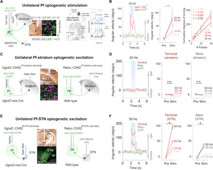
Fig. 2. Unilateral excitation of Pf and Pf-STN Vglut2+ neurons initiates movement and causes ipsiversive turning, but Pf-STR does not.

(A) Optic fiber placement above virally infected Pf Vglut2+ neurons (left). Motion capture during optogenetic stimulation (right). (B) Unilateral excitation (n = 6) (left) increased ipsiversive angular velocity [n = 6, two-way repeated measures analysis of variance (RM ANOVA), before versus after stimulation (F1,5 = 12.46, P = 0.017, n = 6)]. Cumulative ipsiversive rotations in degrees (right) increased as a function of the number of ChR2 excitation pulses (all r2 ≥ 0.98). (C) Illustration of thalamostriatal (Pf-striatum) optogenetic experimental strategies during 3D motion capture. (D) No significant changes in angular velocity were observed during Pf-striatum cell body or terminal ChR2 excitation (two-way RM ANOVA, P > 0.05, n = 8; control mice, n = 6). (E) Schematic showing Pf-STN pathway–specific terminal stimulation and cell body stimulation. (F) Excitation of Pf-STN cell bodies and terminals caused a significant increase in ipsiversive angular velocity (two-way RM ANOVA, Pf-STN cell body: F1,6 = 26.76, P = 0.0021, n = 7; Pf-STN terminal: F1,7 = 62.24, P < 0.0001, n = 8; control mice, n = 6). Error bars indicate SEMs. n.s., not significant. eYFP, enhanced yellow fluorescent protein.
