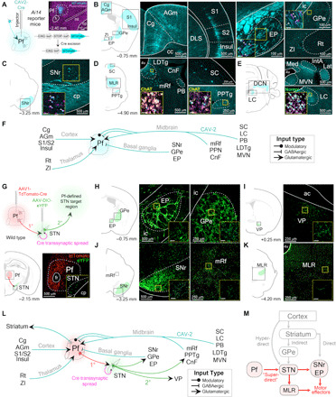
Fig. 6. Viral retrograde tracing reveals that the Pf connectivity with BG and brainstem locomotor regions.

(A) Representative example of CAV2-Cre injection into the Pf of an Ai14 transgenic mouse to visualize afferent projections. (B to E) Retrograde labeled cells from Pf CAV2-Cre injection shown in (A). Cells were found in agranular medial cortex (AGm), Cg, entopeduncular nucleus (EP; rodent homolog of primate internal segment of the globus pallidus), external segment of the globus pallidus (GPe), insular cortex (Insul), reticular nucleus (Rt), S1, S2, and zona incerta (ZI) (B); SNr (C); cuneiform nucleus (CnF), laterodorsal tegmental nucleus (LDTg), mesencephalic reticular formation (mRf), parabrachial nucleus (PB), PPN, and superior colliculus (SC) (D); interposed anterior nucleus of the cerebellum (IntA), lateral cerebellar nucleus (Lat), locus coeruleus (LC), medial cerebellar nucleus (Med), and medial vestibular nucleus (MVN) (E). Insets show retrograde labeled cells colocalized with neuromodulatory cell type markers [choline acetyltransferase (ChAT) and norepinephrine (Norepi)]. (F) Summary schematic of viral-based retrograde Pf anatomy results. (G) Top: Injection of tdTomato-Cre into Pf (first-order neuron) allows transsynaptic spread of Cre into the STN. AAV-DIO-eYFP was subsequently injected into the STN (second-order neuron) to visualize Pf-defined subthalamic projections. Bottom: Illustration and histological confirmation showing Pf and STN injections. (H to K) Coronal sections show terminal labeling sites of Pf-defined subthalamic targets. (L) Schematic of viral-based retrograde Pf and transsynaptic Pf-STN anatomy results. (M) Schematic of the superdirect pathway. ac, anterior commissure; cc, corpus callosum; DCN, deep cerebellar nuclei; ml, medial lemniscus; MLR, midbrain locomotor region.
