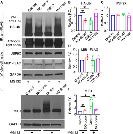
Fig. 4. S-nitrosylation activates USP9X to deubiquitinate MIB1.

HEK293 cells expressing MIB1-FLAG and HA-Ub were cultured in the presence of DETA NONOate (NO donor), GSNO (S-nitrosylating agent), or WP1130 (USP9X inhibitor). (A) After addition of MG132 and immunoprecipitation (IP) with anti-FLAG antibody, immunoblot with anti-HA antibody shows HA-Ub-MIB1-FLAG of various molecular weights in untreated condition (control) and with WP1130, indicating the presence of poly-Ub-MIB1. This is decreased with NO donor and GSNO and is quantified in (B). (C and D) Quantification of USP9X and MIB1-FLAG expression after normalization against GAPDH. (E) pAVICs were cultured in the presence and absence of NO donor for 5 days, and immunoblot for MIB1 is shown with and without the addition of MG132. (F) Quantification of MIB1 is shown after normalization against GAPDH. *P ≤ 0.05. For Western blots (WB), two-tailed unpaired t test was performed. F.C., fold change.
