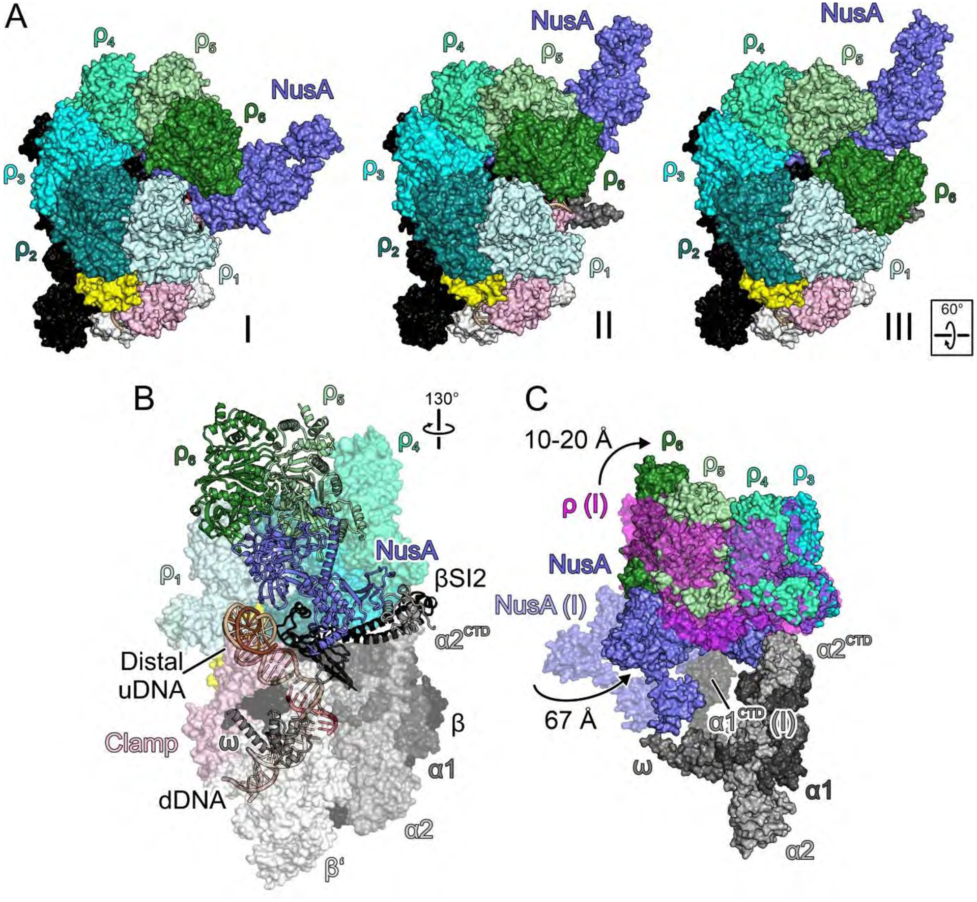
Fig. 3. Priming.

(A) Surface views of the engagement (I), primed (II) and RNA capture (III) complexes, illustrating rotation of NusA underneath ρ6 (I to II) and shift of ρ6 from ρ5 to ρ1 (II to III). (B) Semi-transparent surface/cartoon representations of the primed complex, highlighting contact sites of ρ subunits and distal uDNA on top of β’ZBD. (C) Overlay of selected elements of the primed complex (solid surfaces) and engagement complex (semi-transparent surfaces; ρ, magenta), highlighting movements of NusA and ρ, and handover of NusANTD from α1CTD to α2CTD.
