. 2021 Jan 13;10:e63102. doi: 10.7554/eLife.63102

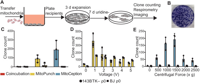
Figure 3. Stable retention of transplanted mitochondrial DNA (mtDNA) into transformed and replication-limited cells.

(A) Workflow for stable isolated mitochondrial recipient (SIMR) cell generation by mitochondrial transfer into ρ0 cells. (B) Representative fixed and crystal violet stained 10 cm plate image following MitoPunch and SIMR cell selection used for SIMR clone generation quantification. (C) Quantification of crystal violet stained 143BTK– ρ0 and BJ ρ0 SIMR clones. Error bars represent SD of three technical replicates. (D) Quantification of crystal violet stained 143BTK– ρ0 and BJ ρ0 SIMR clones formed by MitoPunch actuated with indicated voltages after uridine-free selection. Error bars represent SD of three technical replicates with the exception of BJ ρ0 5 V transfer, which shows two replicates. (E) Quantification of crystal violet stained 143BTK– ρ0 and BJ ρ0 SIMR clones formed by MitoCeption with indicated centripetal forces after uridine-free selection. Error bars represent SD of three technical replicates.

Figure 3.

Figure 3—figure supplement 1. Verification of surviving mitochondrial donor cells following mitochondrial isolation.

Figure 3—figure supplement 1.

Images of three crystal violet stained 10 cm plates seeded with isolated mitochondria from ~1.5 × 10⁷ HEK293T dsRed donor cells taken from three independent mitochondrial isolations following dialyzed medium selection. Pictures were taken on a circular white disk matted within a cardboard frame for clarity.
Figure 3—figure supplement 2. MitoPunch generates stable isolated mitochondrial recipient (SIMR) clones in immortalized mouse cells.

Figure 3—figure supplement 2.

Quantification of crystal violet stained B16 ρ0 SIMR clones formed by MitoPunch transfer of isolated L929 mitochondria actuated with indicated voltages after SIMR cell selection. Error bars indicate the range between the technical duplicates.
Figure 3—figure supplement 3. Quantification of MitoPunch reproducibility.

Figure 3—figure supplement 3.

Quantification of crystal violet stained 143BTK– ρ0 stable isolated mitochondrial recipient (SIMR) clones generated in technical triplicate from three independent HEK293T dsRed mitochondrial donor cell preparations plotted alongside technical singlet DPBS delivery and plated mitochondrial preparation controls (Figure 3—figure supplement 1).
Figure 3—figure supplement 4. Quantification of MitoPunch reproducibility relative to mitochondrial mass transferred.
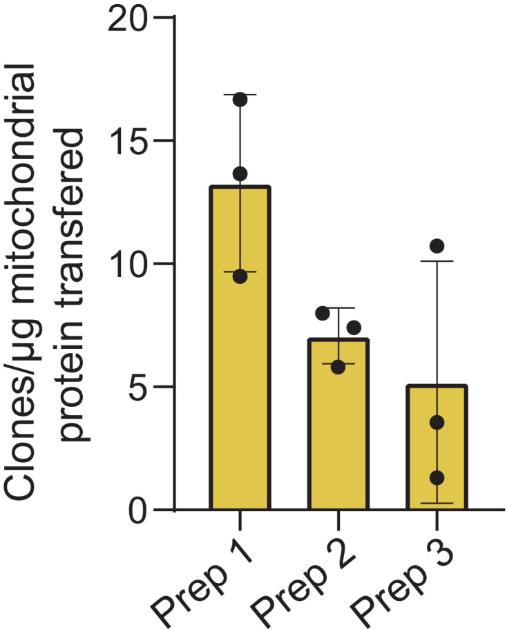
Figure 3—figure supplement 4.

Quantification of crystal violet stained 143BTK– ρ0 stable isolated mitochondrial recipient (SIMR) clones generated in technical triplicate from three independent HEK293T dsRed mitochondrial donor cell preparations plotted as number of SIMR clones generated per µg isolated mitochondrial protein loaded into the polydimethylsiloxane (PDMS) reservoir.
The mass of isolated mitochondria per 120 µL of isolated mitochondrial suspension for the three preparations are as follows: Prep 1–27 µg, Prep 2–33 µg, Prep 3–25 µg.
Figure 3—figure supplement 5. Quantification of stable isolated mitochondrial recipient (SIMR) generation efficiency by delivering different masses of isolated mitochondria.

Figure 3—figure supplement 5.

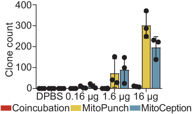
Quantification of crystal violet stained 143BTK– ρ0 SIMR clones using indicated concentrations of mitochondrial suspension following 7 days of culture in SIMR selection medium. Error bars represent SD of three technical replicates.
Figure 3—figure supplement 6. Quantification of MitoPunch stable isolated mitochondrial recipient (SIMR) generation by serial deliveries using one isolated mitochondrial aliquot.

Figure 3—figure supplement 6.

Quantification of crystal violet stained SIMR clones formed by serial MitoPunch deliveries of HEK293T dsRed mitochondria into 143BTK– ρ0 recipient cells using the same used mitochondrial sample remaining in the polydimethylsiloxane (PDMS) reservoir after the preceding delivery.