. 2021 Jan 22;10:e62117. doi: 10.7554/eLife.62117

Figure 5. The L1(atp) → L2(adp) → C1(atp) transition.

(A) A back view of the Mfd-elongation complex (EC) structures is shown, viewing down the axis of the upstream duplex DNA (the direction of transcription would be to the right). The RNA polymerase (RNAP) is shown as a molecular surface with nucleic acids shown in cartoon format. Mfd is shown as transparent molecular surfaces surrounding the backbone ribbon. The bottom row shows the transition through the experimental structures [L1(atp) → L2(adp) → C1(ATP)]. Below that is denoted the translations and rotations of the Mfd domains associated with each transition (superimposed on the EC structure). The large translation/rotation of Mfd D5(TD1)-D6(TD2)-D7 in the L1(atp) → L2(adp) transition must occur by clockwise corkscrewing around the DNA minor groove in order to leave behind the unfolded portion of the RH, which wraps around the DNA. The configuration of Mfd D1–D3 in L1(atp) would block this transition and also result in the entanglement of linkers; thus, we propose that this transition is facilitated by transient intermediates [L1.5a] and [L1.5b], which have been modeled with displaced Mfd D1–D3 tethered by the long linker connecting D3 with the D4(RID). This allows the unencumbered transition of Mfd D5(TD1)-D6(TD2)-D7 from [L1.5a] to [L1.5b] (illustrated by the thick yellow arrow). Mfd D1-D3 then accommodates in a new configuration in L2(adp). The L2(adp) → C1(ATP) transition involves another large translation and rotation of D1–D3 (denoted), which finally exposes the Mfd-D2 UvrA-interacting surface. This transition is also illustrated in Figure 2—video 1.

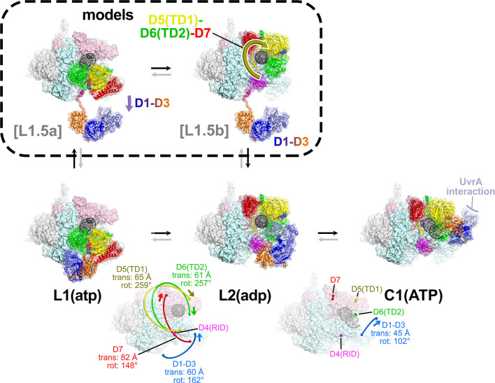
Figure 5.

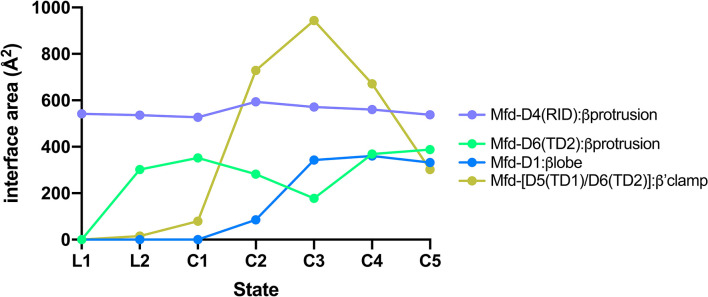
Figure 5—figure supplement 1. Selected interdomain Mfd:RNAP interface areas.

Figure 5—figure supplement 1.

Interface areas [calculated using the PDBePISA server; (Krissinel and Henrick, 2007)] for selected Mfd:RNAP interdomain interfaces, plotted for each Mfd-elongation complex (EC) structural state. Mfd-D4(RID):βprotrusion: The initial engagement of Mfd with the RNA polymerase (RNAP) is through the Mfd-D4(RID):βprotrusion interaction (average interface area of 553 Å2) and this interaction is maintained through all seven states (L1, L2, and C1–C5). Mfd-[D6(TD2)]:βprotrusion: In L1, the Mfd translocation module [D5(TD1)/D6(TD2)] interacts with upstream DNA (roughly −38 to −27) and does not interact with RNAP (Figure 4—figure supplement 1B). Upon the transition to L2, the translocation module walks on the DNA, moving toward the RNAP until it 'bumps' into the RNAP, characterized primarily by D6(TD2) interacting with the RNAP βprotrusion [the Mfd-D4(RID) and D6(TD2) interfaces with the RNAP βprotrusion do not overlap]. Once Mfd-D6(TD2) pushes up against the RNAP βprotrusion in L2, that interface is also maintained throughout the rest of the structures (L2, C1–C5, average interface area of 312 Å2). During the Mfd-nucleotide hydrolysis cycle, the movements of Mfd relative to the RNAP cause cyclical RNAP conformational changes involving RNAP clamp and βlobe-Si1 motions (Figure 7). The RNAP clamp is opened in C3 as Mfd wedges itself between the βprotrusion and the clamp, pushing on the clamp through a large interface with the translocation module (maximum Mfd-[D5(TD1)/D6(TD2)]:β'clamp interface area of 944 Å2 in C3). The RNAP βlobe-Si1 is pushed sideways by an interaction with Mfd-D1 (can be seen in Figure 7).