. 2021 Feb 5;10:e60263. doi: 10.7554/eLife.60263

Figure 2. Lipid II is synthesized in the IMD and trafficked to the PM-CW.

(A) Left, conventional microscopy of M. smegmatis coexpressing PonA1-mRFP and MurG-Dendra2 treated +/- benzyl alcohol (BA). Right, fluorescence distribution of the fusion proteins. a.u., arbitrary units. r denotes the Pearson’s correlation value. 42>n>57. (B) Top, detection of lipid-linked peptidoglycan (PG) precursors from membrane fractions. Bottom, metabolic labeling of mycobacterial cell wall synthesis (García-Heredia et al., 2018). (C) PG precursors are labeled as in (B), top. The labeled precursors are in the IMD and PM-CW of wild-type M. smegmatis but accumulate in the IMD upon MurJ depletion (García-Heredia et al., 2018). While we do not yet understand the loss of signal from fraction 10, we note that there are precursors present but in low abundance (see Figure 2—figure supplement 3B). (D) M. smegmatis-expressing MurG-Dendra2 were incubated with alkDADA. Surface-exposed alkynes on fixed cells were detected by CuAAC (García-Heredia et al., 2018). Bacteria were imaged by SIM-E.

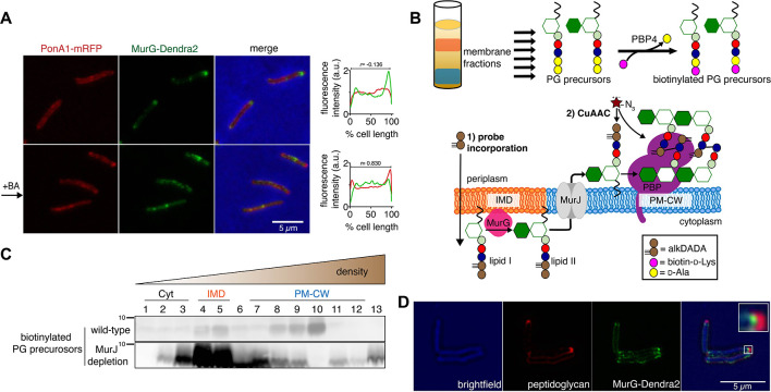
Figure 2.

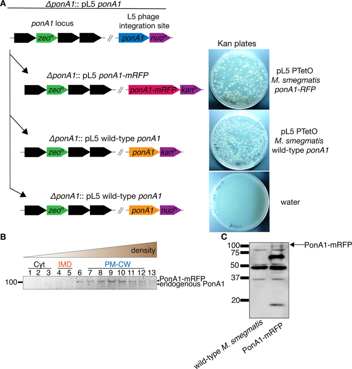
Figure 2—figure supplement 1. PonA1-mRFP is functional.

Figure 2—figure supplement 1.

(A) A plasmid bearing ponA1-mRFP supports the growth of ∆ponA1. In M. smegmatis, ponA1 is essential for viability. ∆ponA1 M. smegmatis complemented with a wild-type copy of ponA1 in the L5 phage integration site (∆ponA1::pL5 wild-type ponA1, Kieser et al., 2015; Baranowski et al., 2018) was transformed with an integrating plasmid bearing either ponA1-mRFP (top right), wild-type ponA1 (center right), or nothing (water; bottom right). Transformants were then selected on kanamycin plates. The kanR ponA1 and kanR ponA1-mRFP cassettes replaced nuoR ponA1 at similar frequencies (not shown), indicating that the fluorescent protein fusion construct can function as the sole copy of ponA1. (B) PonA1-mRFP is active in PM-CW. Lysates from PonA1-mRFP-expressing M. smegmatis were separated by density gradient as in Figure 1B, then incubated with Bocillin-FL as in Figure 1C to label active transpeptidases. The top band corresponds to PonA1-mRFP, and the band immediately below to endogenous PonA1 (see Figure 1—figure supplement 6). (C) Lysates from wild-type and PonA1-mRFP-expressing M. smegmatis were blotted with anti-RFP. The presence of bands that are not present in wild-type lysates and do not correspond to full-length PonA1-mRFP suggests that the fusion protein may undergo degradation.

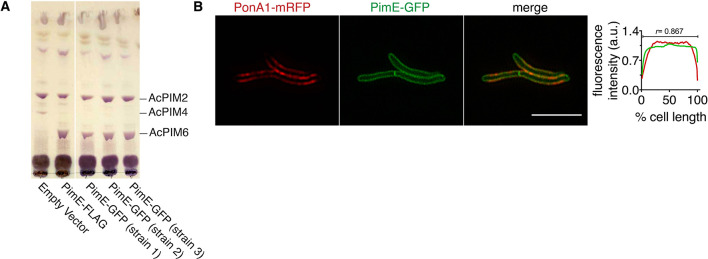
Figure 2—figure supplement 2. PimE-GFP is functional and has a similar subcellular localization to PonA1-mRFP.

Figure 2—figure supplement 2.

(A) PimE is a mannosyltransferase involved in phosphatidylinositol mannoside (PIM) biosynthesis, converting AcPIM4 to more polar PIMs. ∆pimE M. smegmatis was complemented with the indicated expression vectors. Glycolipids were extracted, purified, and separated by thin-layer chromatography. PIMs were visualized by orcinol staining. Similar to PimE-FLAG (Morita et al., 2006), PimE-GFP can restore the production of AcPIM6. (B) PonA1-mRFP distribution overlaps with that of the PM-CW marker PimE-GFP. M. smegmatis coexpressing PonA1-mRFP and PimE-GFP was imaged by SIM-E and conventional microscopy. Left, representative SIM-E image. Scale bars, 5 µm. Right, the fluorescence intensity profiles from conventional microscopy images were quantitated as in Figure 2A. r, Pearson’s correlation value. n = 116 cells.

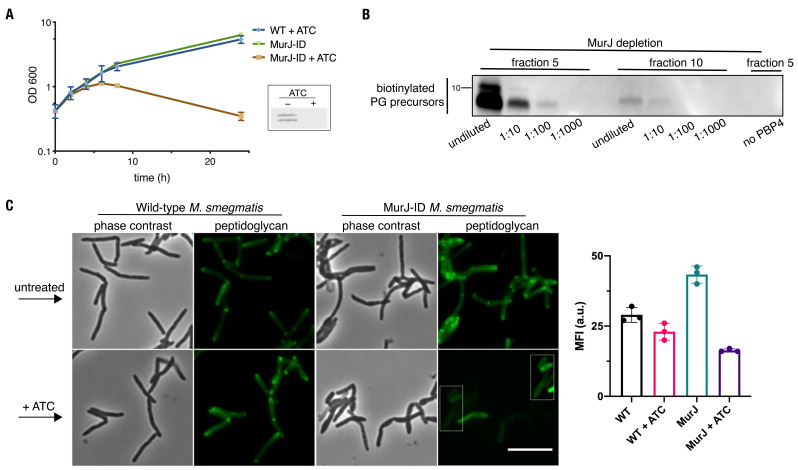
Figure 2—figure supplement 3. MurJ is critical to comparmentalize both vertical and lateral cell wall synthesis.

Figure 2—figure supplement 3.

(A) MurJ is depleted upon treatment with ATC. Depletion strain (MurJ-ID; carrying FLAG tag) was grown +/- ATC. Insert, immunoblot of lysates showing that MurJ is degraded after 8 hr of ATC treatment as reported (Gee et al., 2012). (B) Fractions 5 and 10 from Figure 2C were biotinylated as in Figure 2B, and 10-fold dilutions were made from the biotinylated products. We find substantially more lipid-linked precursors in fraction 5 compared to 10 as in Figure 2C (C) Depletion of MurJ alters amount and location of nascent peptidoglycan. Wild-type (left) or MurJ-ID (depletion strain; right) M. smegmatis in early log phase were treated +/- ATC then incubated with alkDADA. Bacteria were washed, fixed, subjected to CuAAC, and imaged by conventional fluorescence microscopy. Scale bar, 5 µm. Representative experiment (n = 2) performed as in (A) where fluorescence of the peptidoglycan labeling was quantified by flow cytometry. The median fluorescence intensities (MFI) of no probe controls were subtracted from those of the samples. Error bars denote standard deviation of technical triplicates.