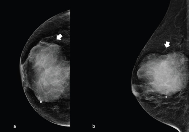
Figure 2. Right side full-field mammography of a 40-year-old patient presenting a unilateral breast mass subsequently identified as a MS. (a): Cranio caudal and (b): mediolateral oblique projection, showing a noncalcified ill-defined radiopaque mass with poorly defined ‘feathery’ margins (arrow).

