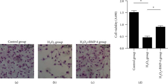
Figure 2.

HE staining of cells. Images (a–c) show that the number of HE-labelled cells decreased under oxidative stress by H2O2 and was markedly increased by the action of BMP-4. (d) Quantification of cell number. Data are expressed as mean ± SEM, ∗p < 0.05.
