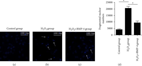
Figure 3.

Hoechst staining of cells. Images (a–c) show alterations in the nuclear morphology after H2O2 and H2O2+BMP-4 treatment of HLE-B3 cells. Arrows indicate the alterations in nuclear morphology. Normal cell nuclei were stained lightly and uniformly. Under oxidative stress, the nuclei were fragmented and stained with dense hyperchromatism. After BMP-4 treatment, the nucleus fragmentation was obviously improved. (d) Quantification of fragmented nuclear number. Data are expressed as mean ± SEM, ∗p < 0.05.
