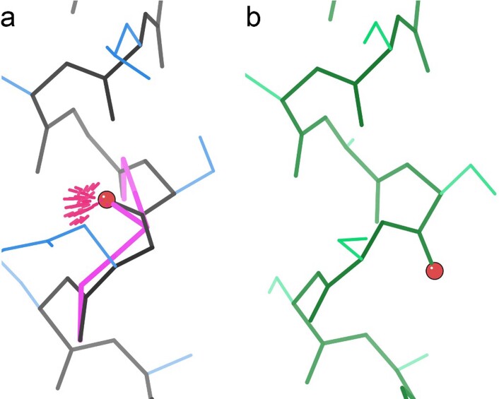
Extended Data Fig. 2. Classic CaBLAM outlier with no Ramachandran outlier.
a, Mis-modeled peptide (identified by red ball at carbonyl oxgen position) is flagged by two successive CaBLAM outliers (magenta dihedrals), a bad clash (hot-pink spikes), and a bond-angle outlier (not shown), but no Ramachandran outlier. b, Correctly modeled peptide, involving a near-180° flip of the central peptide to achieve regular α-helical conformation. Ser 38 of T1/APOF model 60_1 is shown in (a); model 35_1 shown in (b). This example illustrates the most easily correctable situations: (1) for a CaBLAM outlier inside helix or β-sheet, regularize the secondary structure; (2) for two successive CaBLAM outliers, try flipping the central peptide. Molecular graphics were generated using KiNG. Note that sidechains are truncated by graphics clipping.

