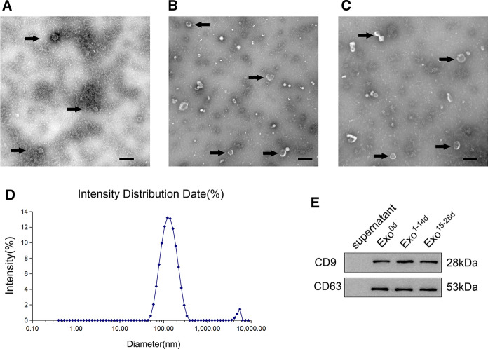
Fig. 2.
Extraction and identification of exosomes derived from human adipose-derived stem cells (hADSCs-Exos) during different time-spans of osteogenesis induction. The representative images of hADSCs-Exos during time-span of osteogenesis induction at Day 0 (Exo0d, A), Day 1 to 14 (Exo1−14d, B), and Day 15 to 28 (Exo15−28d, C) were observed by transmission electron microscopy (TEM). Black arrows indicated the exosomes. Scale bar = 200 nm. D The particle size distribution of hADSCs-Exos was measured by dynamic light scattering (DLS) analysis, and the mean size of hADSCs-Exos was 125.8 ± 3.822 nm. E Western blot analysis showed that the specific surface markers of exosomes (CD9 and CD63) were detected

