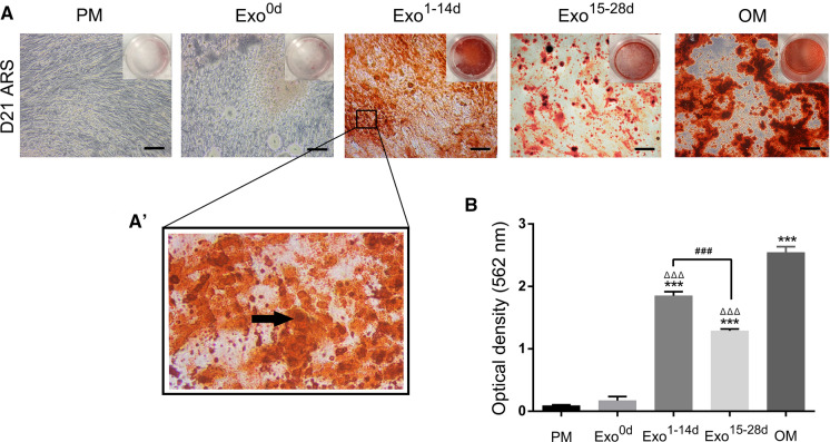
Fig. 4.
Alizarin red S staining (ARS) assays of hADSCs after osteoinduction to determine the optimal time-span for exosomes. A: ARS staining of hADSCs incubated with PM, OM and different time-span of osteogenic induction exosomes (Exo0d, Exo1−14d and Exo15−28d) on day 21. Scale bar = 200 μm. A’ The higher magnification panels of black box in Fig. 4A. The black arrow indicated the calcified nodules. B Quantification of ARS of hADSCs treated with or without exosomes. PM: proliferation medium; OM: osteogenic medium. ***p < 0.001 compared with PM group, ΔΔΔp < 0.001 compared with OM group, ###p < 0.001 represents significant differences between compared groups. (Color figure online)

