Fig. 5.
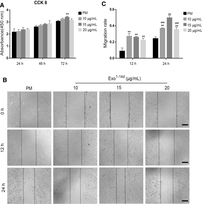
hADSCs-Exos enhanced the proliferation and migration of hADSCs. A CCK-8 analysis of hADSCs after treatment with exosomes (10, 15 and 20 μg/mL) from hADSCs after 14 days of osteogenic differentiation (Exo1−14d) for 24, 48, and 72 h. B, C Migration of hADSCs co-cultured with different concentration of Exo1−14d (10, 15 and 20 μg/mL) assessed by wound healing assay in 0 h, 12 h and 24 h. Scale bar = 200 μm. The quantitative results of wound healing assay.PM: proliferation medium; **p < 0.01 compared with PM group, ###p < 0.001 compared with 15 μg/mL Exo1−14d group

