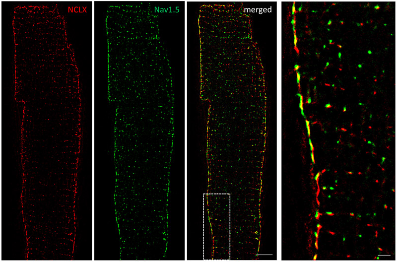
Figure 4. Mitochondrial NCLX is mainly found in SSM near Nav1.5 clusters.
Dual-color stochastic optical reconstruction microscopy (STORM)-acquired images of NCLX (red) and Nav1.5 (green) in single myocytes dissociated from wild-type mice. White-boxed area enlarged in right image. Notice abundance of NCLX signal at lateral membrane and proximity to Nav1.5 clusters. Scale bar 5 μm. Inset scale bar 1 μm.

