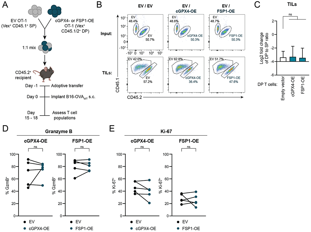
Figure 5. Overexpression of cGPX4 or FSP1 Does Not Affect CD8+ T cell Anti-Tumor Function In Vivo.

A, Schematic depicting adoptive transfer experiment. The mouse image was created using BioRender.com.
B,C, Representative plots (B) and quantification (C) of flow cytometric measurement of the change in ratio of CD45.1/2 DP to CD45.1 SP among Vex+ CD8+ T cells in B16-OVAWT tumors compared to input. Tumors were harvested for analysis on day 15 to 18 after implantation. Ratio fold-change calculated as (DP:SP)TIL / (DP:SP)input. n=5-8 animals per group. Statistical significance determined by one-way ANOVA.
D,E, Percentages of Vex+ CD8+ T cells expressing GzmB (D) and Ki-67 (E) as measured by flow cytometry after tumor harvest. n=5 mice per group. Statistical significance determined by paired t tests.
SP, CD45.1 single-positive. DP, CD45.1/2 double-positive. *p<0.05 **p<0.01 ***p<0.001 ****p<0.0001. ns, not significant. Graphs display mean +/− SD. Data representative of ≥2 independent experiments. TIL, tumor-infiltrating lymphocyte.
