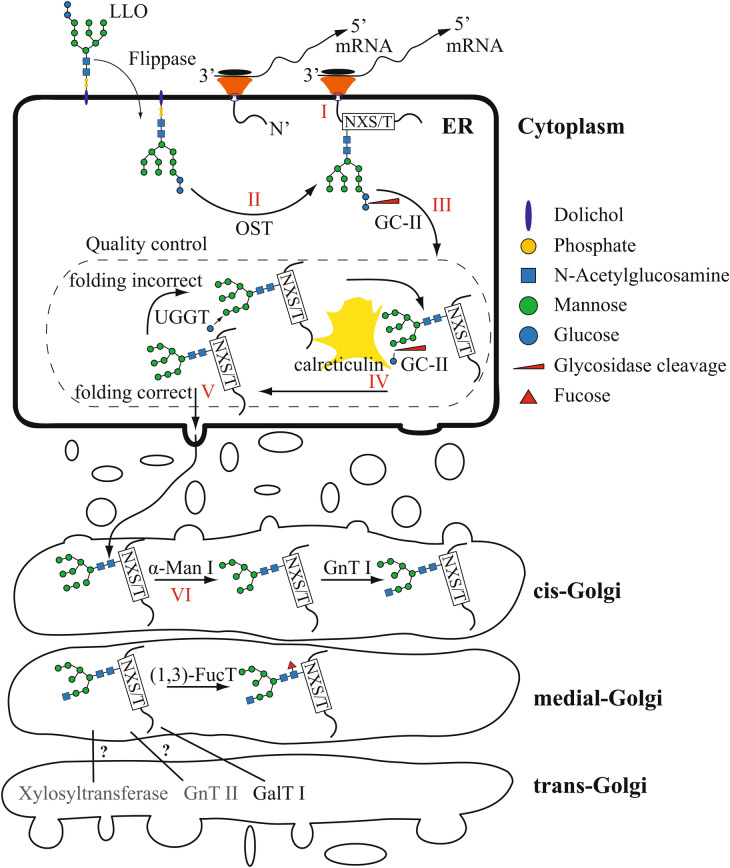
Figure 2.
Simplified scheme of the N-linked glycosylation pathway in the endoplasmic reticulum and the Golgi apparatus. The N-linked glycosylation pathway is shown in a simplified scheme. The mRNA is being attached and translated at the membrane of the endoplasmic reticulum (I), and the glycan is attached by the oligosaccharyltransferase (OST) (II) and trimmed by α-glucosidase II (GC-II) (III). The protein enters the folding control (dashed box) (IV). Calreticulin and UDP-glucose glucosyltransferase (UGGT) are the main component of the system. Correctly folded proteins are further transported to the Golgi apparatus (V), and N-linked glycan structures are further processed by specific enzymes (VI). The following abbreviations are used: alpha-mannosidase (α-Man), N-acetylglucosaminyltransferase (GnTI), 1,3 fucosyltransferase ((1,3)-FucT), galactosyltransferase (GalTI). In grey are two enzymes as examples of missing proteins that are involved in glycan maturation in other organisms. Components of the glycan structure are indicated with blue being dolichol, yellow is phosphate, blue spheres are glucose residues, green spheres are mannose residues, and blue squares are N-acetylglucosamines.

