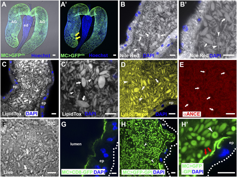
Fig. 1.
The AG lumen contains abundant lipophilic microcarriers. (A and A′) Fluorescence image with (A) and without (A′) bright-field illumination of paired D. melanogaster male AGs connecting to the ejaculatory duct (ed). Main cells express nuclear GFP under Acp26Aa-GAL4 main cell-specific control (green), but secondary cells in distal tip (two of which are marked by yellow arrows in A′) do not. (B–E) Confocal sections through AG lumen stained with Nile Red (B, B′; latter is high magnification view), LipidTox (C and C′), LysoTracker Deep Red (D; yellow) and anti-ANCE (red), a soluble secreted protein (E). White arrows mark representative large microcarriers, and arrowheads mark small microcarriers. (F) DIC image of lumen from living AG also reveals microcarriers (white arrows). (G) Transmembrane CD8-GFP expressed in main cells marks the apical plasma membrane, but not luminal microcarriers. (H and H′) Main cell-expressed GFP-GPI labels microcarriers at their surface (H and H′, white arrowheads) together with the apical surface of the epithelial monolayer (H′, red arrows). Nuclei marked with Hoechst (A and A′, blue) or DAPI (B, E, G, and H, blue). AG epithelium (ep) (dotted white line in G and H marks approximate basal surface). Main cell-specific Acp26Aa-GAL4 driver (MC>). (Scale bars: 10 µm.)

