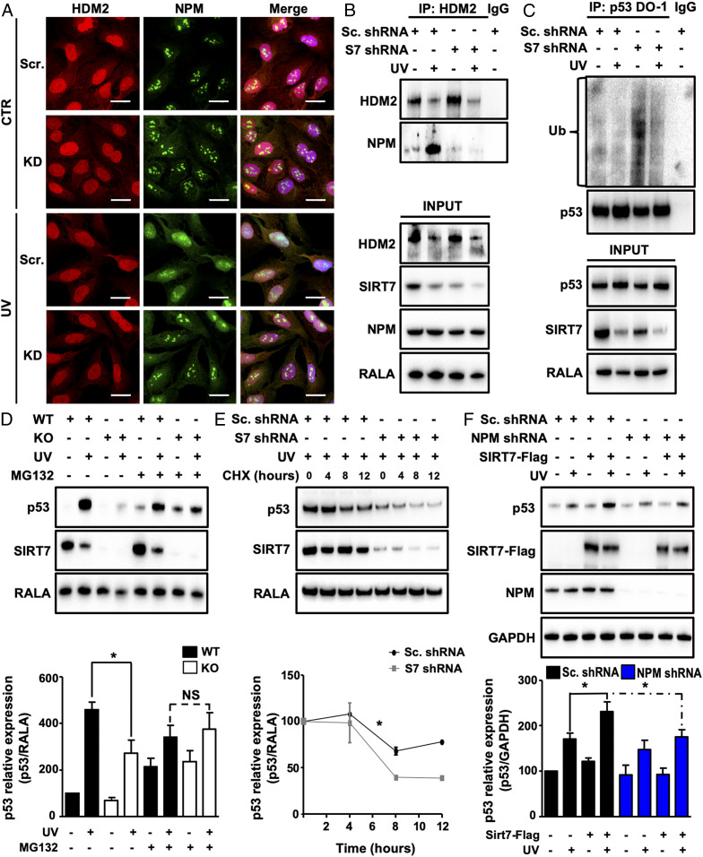
Fig. 3.
SIRT7 enhances association of NPM with HDM2 and prevents p53 ubiquitination under UV. (A) IF staining for NPM and HDM2 of SIRT7 KD and control U2OS cells 12 h after UVC irradiation. Nuclei were counterstained with DAPI (n = 3). (Scale bars, 25 µm.) (B) Coupled immunoprecipitation (IP) (HDM2 antibody) and Western blot analysis (NPM antibody) of SIRT7 KD and control (scrambled) U2OS cell lysates 3 h after UVC irradiation (n = 3). (C) Coupled IP (p53-DO-1 antibody) and Western blot analysis (ubiquitin antibody) of SIRT7 KD and control (scrambled) U2OS cell lysates 2 h after UVC irradiation followed by a 5-h incubation with 10 µM MG-132 (n = 3). The membrane was reprobed with anti-p53 antibody to ensure equal IP of p53 (Top). (D) Western blot analysis of p53 levels in WT and SIRT7 KO MEFs 12 h after UVC irradiation, followed by a 5-h treatment with 10 µM MG-132. RALA was used as loading control. Quantification of p53 relative levels ± SD is given (Bottom) (n = 3). (E) Western blot analysis of p53 levels in control (scrambled) and SIRT7 KD U2OS cell lysates 12 h after UVC irradiation followed by treatment with cycloheximide (CHX; 50 µg/mL). Quantification of p53 relative levels ± SEM is given (Bottom) (n = 3; two-way ANOVA). (F) Western blot analysis of p53, SIRT7, and NPM levels in NPM KD and control (scrambled) U2OS cells transfected with Flag-SIRT7 5 h after UVC irradiation. GAPDH was used as a loading control. Quantification of p53 relative levels ± SD is given (Bottom) (n = 3). *P < 0.05.

