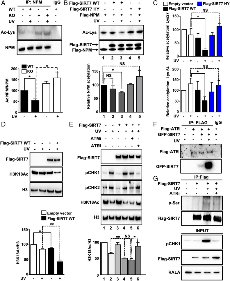
Fig. 4.
UV irradiation stimulates the enzymatic activity of SIRT7 required for efficient deacetylation of NPM. (A) Coupled IP (anti-NPM antibody) and Western blot analysis (anti-acetyllysine antibody) of SIRT7 WT and KO MEFs 5 h after UVC irradiation in the presence of trichostatin A (n = 3). Equal IP of NPM was verified by Western blotting. Quantification of acetylated NPM normalized to immunoprecipitated NPM protein ± SD (Bottom) (n = 4). (B) In vitro deacetylation assay using precipitated Flag-SIRT7 wild type (Flag-SIRT7 WT), SIRT7 catalytically inactive mutant (Flag-SIRT7 HY), and Flag-NPM after overexpression in 293T HEK cells followed by UVC exposure. NPM deacetylation was determined using an anti-acetyllysine antibody (Ac-Lys). Quantification of relative NPM acetylation is shown (Bottom) (n = 3). (C) Relative acetylation levels ± SD of NPM at lysine 27 (Top) and lysine 54 (Bottom) as determined by mass spectrometry of immunoprecipitated NPM using 293T HEK cells transfected with an empty vector, Flag-SIRT7 WT, and Flag-SIRT7 HY 5 h after UVC irradiation (n = 4). (D) Western blot analysis of H3K18Ac in U2OS cells transfected with an empty vector or with Flag-SIRT7 WT 5 h after exposure to UVC. Quantification (Bottom) (n = 3). (E) Western blot analysis of H3K18Ac in 293T HEK cells transfected with an empty vector or with Flag-tagged wild-type SIRT7 (Flag-SIRT7) after exposure to UVC. Cells were pretreated with vehicle, ATR inhibitor (ATRi; 4 µM), and ATM inhibitor (ATMi; 10 µM) prior to exposure to 40 J/m2 UVC, cultivated for 5 h with inhibitors, and analyzed by Western blot. Inhibition of ATR and ATM was assessed by determining phosphorylation levels (p) of CHK1 and CHK2. Quantification of H3K18Ac level normalized to total histone 3 (H3) is shown (Bottom) (n = 3). (F) Coupled IP (anti-Flag antibody) and Western blot analysis (anti-Flag and anti-GFP antibodies) of 293T HEK cells transfected with GFP-SIRT7 and Flag-ATR 5 h after UVC irradiation (n = 3). (G) Coupled IP (anti-Flag M2 affinity beads) and Western blot analysis (anti-phosphorylated serine and anti-Flag antibodies) of 293T HEK cells treated with vehicle or ATRi 5 h after UVC irradiation (n = 3). *P < 0.05, **P < 0.01.

