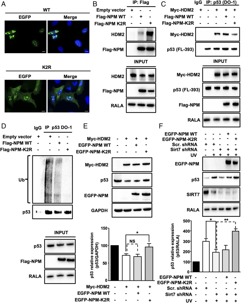
Fig. 5.
SIRT7-mediated deacetylation of NPM at lysines 27 and 54 promotes p53 stabilization by inhibiting HDM2. (A) IF staining of NPM KD U2OS cells transfected with WT EGFP-NPM and K2R EGFP-NPM using an anti-EGFP antibody. Cell nuclei were counterstained with DAPI (n = 3). (Scale bars, 10 µm.) (B) Coupled IP (anti-Flag M2 affinity beads) and Western blot analysis (antibody against endogenous HDM2) of 293T HEK cells transfected with empty vector, Flag-NPM WT, and Flag-NPM K2R mutant. Inputs are shown (Bottom) (n = 3). (C) Coupled IP (p53-DO-1 antibody) and Western blot analysis (antibody against Myc-HDM2) of 293T HEK cells transfected with Myc-HDM2, Flag-tagged WT, and K2R NPM followed by a 5-h treatment with MG-132. p53 concentrations were assessed using an anti-p53 antibody. Inputs are shown (Bottom) (n = 3). (D) Coupled IP (p53-DO-1 antibody) and Western blot analysis (anti-ubiquitin antibody) of U2OS cells transfected with empty vector, Flag-tagged WT and K2R NPM followed by a 5-h treatment with 10 µM MG-132. Inputs are shown (Bottom) (n = 3). (E) Western blot analysis of p53 levels in U2OS cells transfected with empty vector, Myc-tagged HDM2, Flag-tagged WT, and K2R NPM. GAPDH was used as a loading control. The graph represents the average of relative p53 levels ± SD (n = 4). (F) Western blot analysis of p53 in scrambled and SIRT7 KD cells transfected with EGFP-tagged WT and K2R NPM following UVC irradiation 48 h posttransfection and harvested 5 h after irradiation (n = 4). *P < 0.05, **P < 0.01.

