Fig. 5.
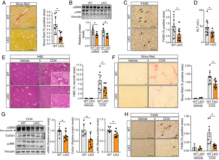
Loss of miR-33 in liver protects against HFD- and CCl4-induced hepatic fibrosis. Characterization of factors related to fibrosis, inflammation, and liver function in male miR-33loxP/loxP (WT) and miR-33loxP/loxP/AlbuminCre (LKO) mice. (A) Representative images and quantification of Sirius red staining in liver sections from WT and LKO mice fed an HFD (n = 9 to 10). (B) Representative Western blots and densitometric analysis of αSMA, pJNK, and housekeeping standard Vinculin in WT and LKO mice fed an HFD (n = 5). (C) Representative images and quantification of F4/80 staining in liver sections from WT and LKO mice fed an HFD (n = 9 to 10). Arrows indicate F4/80-positive cells. (D) Serum ALT levels in WT and LKO mice fed an HFD (n = 8 to 10). (E) Representative images of H&E staining in liver sections from WT and LKO mice treated with vehicle or CCl4 for 6 wk. (Insets) Increased-magnification views of the presence or lack of necrotic areas, which are also quantified (Right; vehicle, n = 3; CCl4, n = 8). (F) Representative images and quantification of Sirius red staining in liver sections from WT and LKO mice treated with vehicle or CCl4 for 6 wk (vehicle, n = 2; CCl4, n = 8). (G) Representative Western blots and densitometric analysis of αSMA, Col3a1, pJNK, and housekeeping standard Vinculin in WT and LKO mice treated with CCl4 for 6 wk (n = 7 to 8). (H) Representative images and quantification of F4/80 staining in liver sections from WT and LKO mice treated with vehicle or CCl4 for 6 wk (vehicle, n = 3; CCl4, n = 8). Arrows indicate F4/80-positive cells. Data represent the mean ± SEM (*P ≤ 0.05 compared with WT animals).

