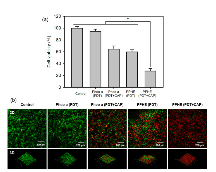
Figure 8.
(a) In vitro cytotoxicity of the PDT and CAP combination therapy on the 3D cell culture model of the CaSki cells evaluated by the MTT assay (n = 4). * p < 0.05 for comparison between two treatment groups. (b) Fluorescence microscopy images of the 3D cell culture model of the CaSki cells after treatment with free Pheo a and PPHE polymeric nanoparticles (4 μg/mL Pheo a), followed by irradiation using a 671 nm laser (42 mW/cm2, 5 min) and CAP (exposure time = 50 s). Live and dead cells were stained with calcein-AM (green) and EthD-1 (red), respectively.

