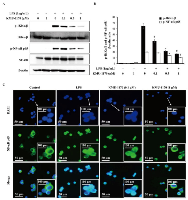
Figure 4.

Inhibitory effect of KMU-1170 on LPS-induced phosphorylation of IKKα/β and NF-κB, and nuclear translocation of NF-κB in THP-1 cells. Cells were differentiated into macrophages for 24 h using PMA (100 nM). And then the cells were treated with LPS (1 μg/mL) for 6 h after pretreatment with the indicated concentrations of KMU-1170 for 1 h. (A) Whole cell lysates were isolated and used to measure the protein expression levels of p-IKKα/β, IKKα/β, p-NF-κB p65, NF-κB p65, and β-actin by Western blot analysis. (B) Image-J software was used to analyze the relative optical density of the p-IKKα/β and p-NF-κB p65 band. (C) Cells were stained with antibodies to the NF-κB p65 (green) and 4′,6-diamidino-2-phenylindole (blue) and captured at ×200 using fluorescence microscope. Arrows indicate 400 times magnification. * p < 0.01 and # p < 0.001 compared to LPS alone.
