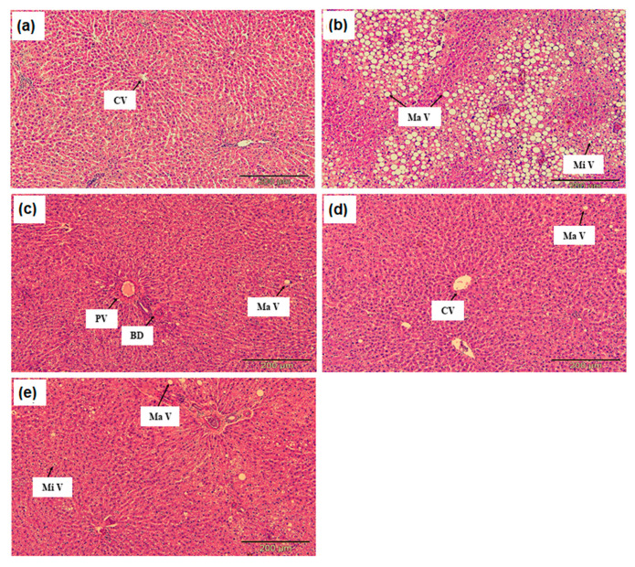
Figure 1.
(a) Histology section of liver tissue in the normal rat group (NG), (b) hypercholesterolemic positive control rats group (PG), (c) hypercholesterolemic rats treated with 2% DPO (HG), (d) hypercholesterolemic rats treated with 2% DDP (DG) (e) hypercholesterolemic rats treated with 10 mg/kg simvastatin (SG) observed under a light microscope at 200× magnification (hematoxylin and eosin staining). CV: central vein; MaV: macrovesicular steatosis; MiV: microvesicular steatosis; PV: portal vein; BD: bile duct.

