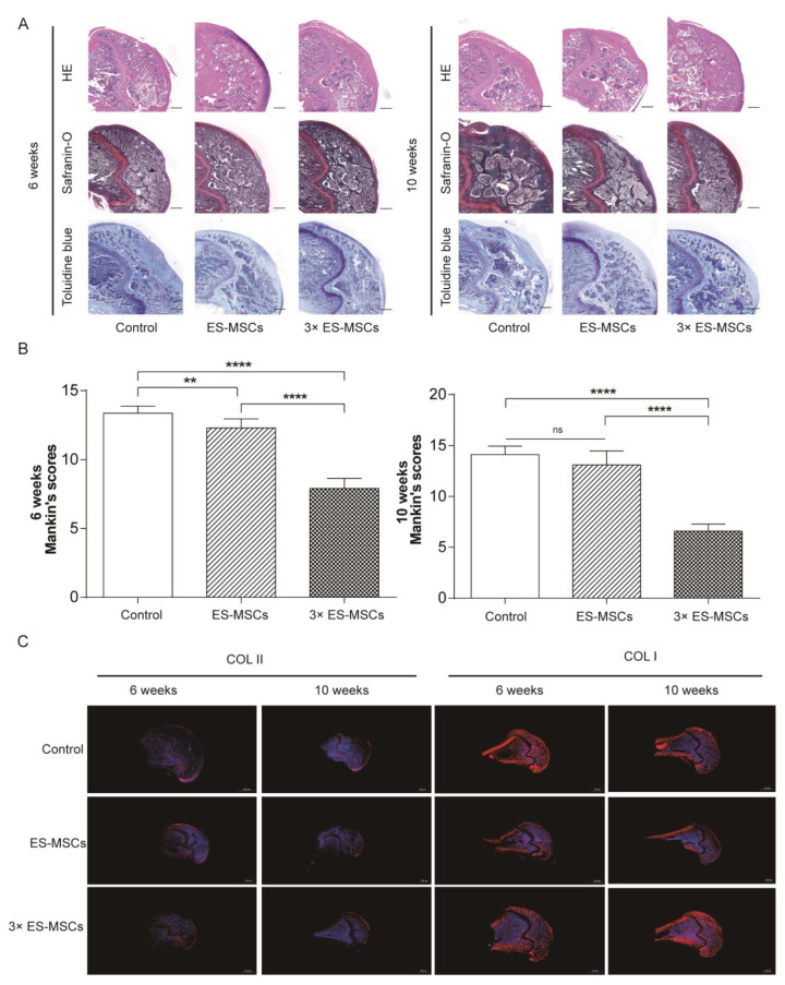
Figure 4.
Histological analysis of the knee joint. (A) Histological images showing HE, safranin O and toluidine blue staining of the medial femoral condyle in all groups, at 6 and 10 weeks after the first injection. (B) The corresponding modified Mankin scoresfor all groups. (C) Immunofluorescence staining for collagen types I and II in all groups at 6 and 10 weeks after the first injection.Error bars represent 95% confidence intervals (CI).** p < 0.01, **** p < 0.001, ns = no significance.

