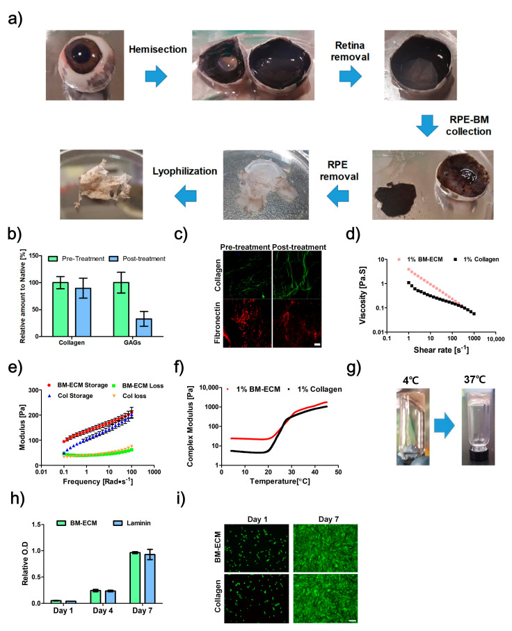
Figure 1.
The development process of Bruch’s membrane-derived extracellular matrix bioink (BM-ECM). (a) Isolation procedure for BM. (b) Biochemical analysis of collagen and glycosaminoglycans (GAGs) for BM pre- and post-treatment with sodium dodecyl sulfate (SDS). (c) Immunofluorescence images of collagen type-1 and fibronectin for BM pre- and post-treatment with SDS. Rheological analysis results, (d) Viscosity, (e) storage modulus and loss modulus, and (f) complex modulus of BM-ECM and collagen in different conditions. (g) Sol-gel transition of 1% BM-ECM as a result of an increase in temperature. (h) Proliferation of ARPE-19 on non-coated, collagen, and BM-ECM-coated surfaces. (i) Live/Dead assay of ARPE-19 on non-coated, collagen, and BM-ECM-coated surfaces (Green: Live, Red: Dead). Scale bar: (c) 50 μm, (i) 200 μm. The error bars represent the standard deviation.

