Figure 3.
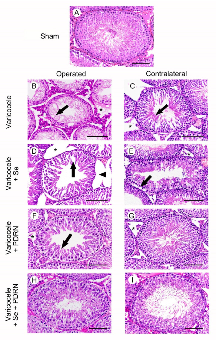
Structural organization of testes from rats of sham, varicocele, varicocele contralateral, varicocele plus Se (0.4 mg/kg/day i.p.), varicocele plus Se contralateral, varicocele plus PDRN (8 mg/kg/day i.p.), varicocele contralateral plus PDRN, varicocele plus Se plus PDRN, varicocele contralateral plus Se plus PDRN groups. (A): In sham testes, the seminiferous tubules have normal morphology. (B): Varicocele rats. The seminiferous epithelium shows reduced thickness with spermatogonia arranged on 1–2 rows and residual condensed sperm tails (arrow). In the extratubular compartment, a marked edema is evident (*). (C): Contralateral testes of the same group. The seminiferous tubules show many elongated spermatids (arrow). The extratubular compartment is edematous and hyperemic (*). (D): In varicocele operated rats treated with Se, the seminiferous epithelium is detached from the basement membrane (arrowhead) and many large clefts are present in its wall (arrow). The extratubular compartment is enlarged (*). (E): In the contralateral testes of the same group, peripheral spermatogonia are often detached from the basement membrane (arrow). The extratubular compartment shows hyperemic blood vessels and edema (*). (F): Varicocele operated rats treated with PDRN. The seminiferous epithelium shows intercellular clefts among round or elongated spermatids (arrow). A mild edema is present in the extratubular compartment (*). (G): Contralateral testes of the same group. The seminiferous epithelium has many elongated spermatids and mature spermatozoa; the extratubular compartment shows only a mild edema (*). (H,I): In the seminiferous tubules of both operated and contralateral testes of varicocele rats treated with Se plus PDRN, the seminiferous epithelium is close to normal. (Scale bar = 50 µm).

