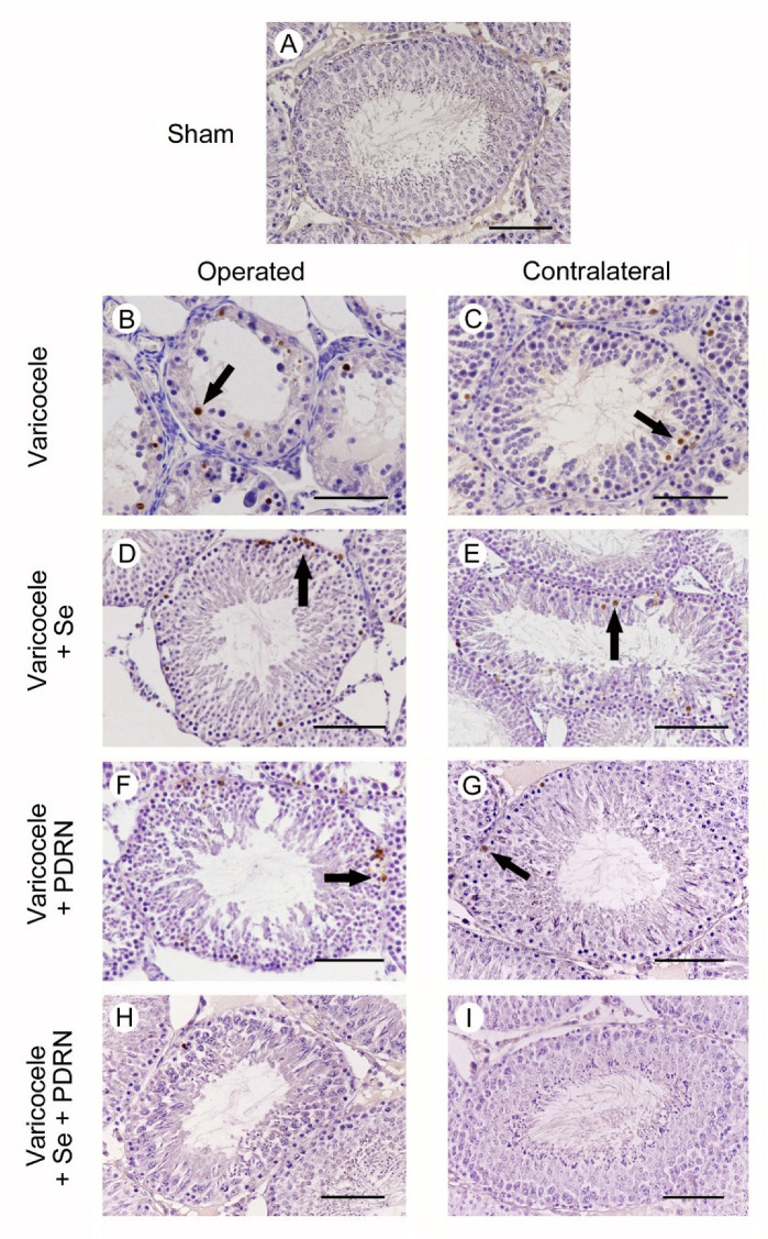
Figure 4.
Assessment of apoptosis in the testes with TUNEL staining technique from rats of sham, varicocele, varicocele contralateral, varicocele plus Se (0.4 mg/kg/day i.p.), varicocele plus Se contralateral, varicocele plus PDRN (8 mg/kg/day i.p.), varicocele contralateral plus PDRN, varicocele plus Se plus PDRN, varicocele contralateral plus Se plus PDRN groups. (A): In sham rats, no TUNEL positive cells can be observed. (B): In the testes of varicocele rats, a large number of TUNEL-positive germ cells (arrow), placed along the wall of the tubules, are observed. (C): In the contralateral testes of the same rats, some isolated TUNEL-positive germ cells are present in the peripheral part of the seminiferous tubules (arrow). (D): In varicocele rats treated with Se, many TUNEL-positive cells are present in the periphery of the seminiferous tubules (arrow). (E): In the contralateral testes of the same group, some TUNEL-positive cells are located in the external part of the tubules (arrow). (F): In the testes of varicocele rats treated with PDRN, the number of TUNEL-positive cells is reduced, and they are located along the peripheral part of the tubules (arrow). (G): In contralateral testes of the same group, only a few TUNEL-positive cells (arrow) are present in the seminiferous epithelium. H, I: In the seminiferous tubules of both operated and contralateral testes of varicocele rats treated with Se plus PDRN, very rare or no TUNEL-positive cells are observed. (Scale bar: 50 µm).

