Figure 6.
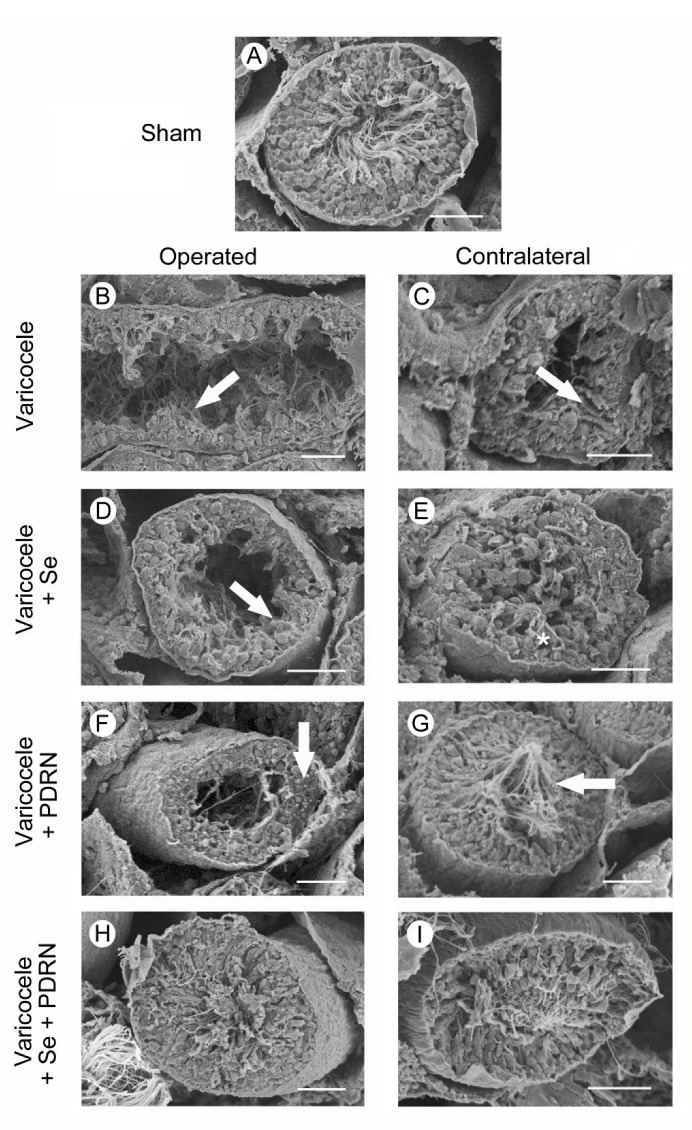
Scanning electron micrographs of testes from sham, varicocele, varicocele contralateral, varicocele plus Se (0.4 mg/kg/day i.p.), varicocele plus Se contralateral, varicocele plus PDRN (8 mg/kg/day i.p.), varicocele contralateral plus PDRN, varicocele plus Se plus PDRN, varicocele contralateral plus Se plus PDRN groups. (A): Sham animals. Note the normal structure of the seminiferous tubules. (B): Varicocele rats. Tubules show evident reduction in their height and condensed sperm tails (arrow). (C): In the contralateral testes of the same group, some clefts are present in the seminiferous epithelium (arrow). (D): In varicocele rats treated with Se, a low and irregularly arranged seminiferous epithelium is evident (arrow). (E): In the contralateral testes of the same group, only a few spermatozoa can be observed (asterisk). (F): In varicocele rats treated with PDRN the tubular lumen is reduced, owing to the presence of a higher epithelium (arrow). (G): In the contralateral testes of the same group, many spermatozoa are present (arrow). (H,I): In varicocele rats treated with Se plus PDRN, both the operated and the contralateral testes show close to normal organization. (Scale bar: 50 μm).

