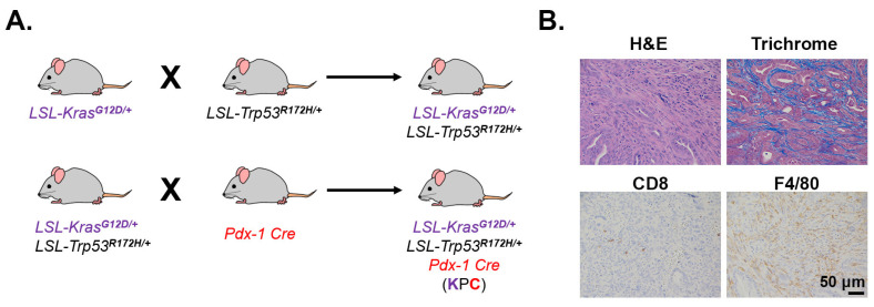
Figure 4.
Desmoplasia, CD8+ T cells, and macrophage infiltration in GEMM KPC tumors. (A). Breeding scheme to generate the genetically engineered KPC mouse model. LSL, LoxP-Stop-LoxP; Pdx-1, pancreatic and duodenal homeobox-1. (B). A PDAC tumor in a 6-month old KPC mouse was H&E or trichrome stained (10×) or stained by immunohistochemistry for CD8+ T cells and the macrophage marker F4/80 (20×). Antibodies used: CD8 (Cell Signaling, #98941) and F4/80 (Cell Signaling, #70076).

