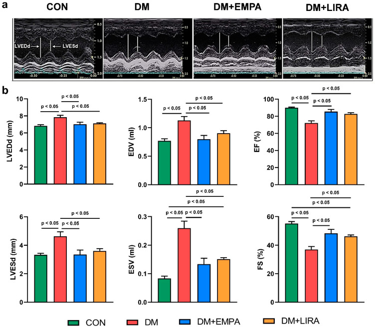
Figure 1.
Echocardiograms of control (CON), diabetes mellitus (DM), empagliflozin-treated DM (DM + EMPA) rats, and liraglutide-treated DM (DM + LIRA) rats at 16 weeks of age. (a) Representative M-mode echocardiograms of CON (N = 8), DM (N = 8), DM + EMPA (N = 8), and DM + LIRA rats (N = 8). (b) Bar graphs of echocardiographic measurement results of left ventricle (LV) end-diastolic and end-systolic diameters (LVEDd and LVEDs), end-diastolic volume (EDV), end-systolic volume (EDV), ejection fraction (EF), and fraction shortening (FS).

