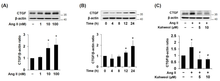
Figure 1.
Effect of kahweol on connective tissue growth factor (CTGF) protein expression in Ang II-stimulated vascular smooth muscle cells (VSMCs). (A,B) Western blot analysis of CTGF in Ang II-treated VSMCs. Cells were exposed to various concentrations of Ang II (0, 1, 10, and 100 nM) for 24 h. In addition, cells were treated with Ang II 100 nM for the indicated times (0, 4, 8, 12, and 24 h). Protein samples were refined from cultured VSMCs treated with Ang II. (C) Cells were pretreated in the presence or absence of kahweol (0, 5, and 10 μM) for one hour, and then stimulated with 100 nM Ang II for a further 24 h. Protein levels of CTGF and β-actin were determined by Western blotting with specific antibodies. Bar graphs present the densitometric quantification of the Western blot bands. Results are representative of three independent experiments. *, p < 0.05 vs. untreated control; #, p < 0.05 vs. Ang II group.

