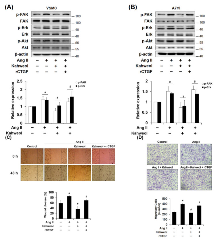
Figure 3.

The involvement of focal adhesion kinase (FAK) and Erk in the mechanism underlying the kahweol effect on migration in Ang II-stimulated VSMCs. (A,B) Cells were pretreated with rCTGF and kahweol at one hour intervals and then stimulated with Ang II for 24 h. Protein levels of phosphorylated FAK, Erk, Akt, and β-actin were determined by Western blotting with specific antibodies. Bar graphs present the densitometric quantification of the Western blot bands. (C) Cells were scratched with a micropipette tip to form a cell- free (wounded) area and pretreated with rCTGF and kahweol at one hour intervals and then stimulated with Ang II for 48 h. Wound areas were visualized using a phase-contrast microscope. The distance migrated was determined as the average of the distances of each cell from the wound boundary. (D) For selective migration assay, a transwell assay was performed. Cells were seeded on the inner chamber under the same treatment condition, as in Figure 3C. After fixing, cells were visualized by crystal violet staining. Unmigrated cells were scraped off, and the migrated cells were counted under a light microscope. Results are representative of three independent experiments. *, p < 0.05 vs. untreated control; #, p < 0.05 vs. Ang II group; §, p < 0.05 vs. kahweol + Ang II treated group.
