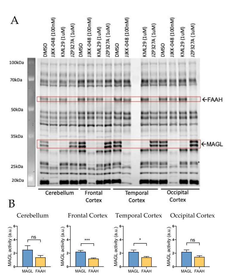
Figure 3.
Competitive gel-based ABPP reveals MAGL and FAAH activity in rat cerebellum and cortex. (A) Cerebellar and frontal, temporal and occipital cortexes proteomes were incubated for 1 h with vehicle (DMSO), MAGL inhibitors JJKK-048 (100 nM) and KML29 (1 μM) and FAAH inhibitor JZP327A (1 μM), and then labeled with the fluorescent probe TAMRA-FP, as indicated in Materials and Methods. FAAH and MAGL were identified based on selective inhibition and their expected molecular weights. Both MAGL and FAAH activities were high in the cerebellum and cortex. (B) Histograms showing the basal activity of MAGL and FAAH in the cerebellum and frontal, temporal and occipital cortexes. Basal MAGL activity was ~2-fold higher compared to that of FAAH in frontal and temporal cortexes. In contrast, MAGL and FAAH activities were not found statistically different in samples of the cerebellum and occipital cortex. Unpaired t-test, * p < 0.05, *** p < 0.001, ns = nonsignificant, n = 10.

