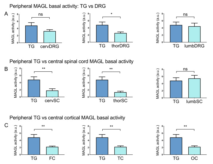
Figure 4.
Comparing the basal MAGL activity in peripheral and central rat tissues. (A) Comparison of basal MAGL activity between TG and DRG. Basal MAGL activity was ~2-fold higher in TG than in thoracic DRG but no significant difference was found in TG vs cervical and lumbar DRG. (B) Comparison of basal MAGL activities between peripheral TG and CNS spinal cord tracts. Basal MAGL activity in the cervical (cervSC) and thoracic (thorSC) spinal cord was ~3-fold lower than in TG. The level of MAGL activity was similar between samples of TG and the lumbar spinal cord (lumbSC). (C) Comparison of basal MAGL activities between peripheral TG and cortical samples. Basal MAGL activity in frontal (FC), temporal (TC) and occipital cortexes (OC) was approximately half of that in the TG sample. Unpaired t-test, * p < 0.05, ** p < 0.01, ns = nonsignificant, n = 8 (TGs, DRGs), n = 11 (BS, cSC), n = 8 (tSC, lSC) and n = 10 (Cbl, cortex).

