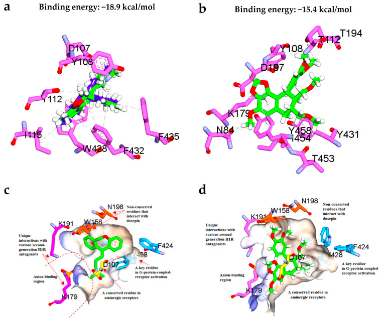
Figure 7.
(a) Superposition of the docked conformation of doxepin and the original cocrystal doxepin retrieved from the protein crystal structure (PDB ID: 3RZE). The original cocrystal doxepin is colored blue while the docked doxepin is colored green for carbon atoms. (b) Binding interactions of the most active compound hypophyllanthin (2) with the adjacent residues in the H1 receptor binding site. (c) Position of the key residues of the H1 receptor binding site in complex with doxepin as retrieved from the protein crystal structure (PDB ID: 3RZE). (d) Position of the key residues of the H1 receptor binding site in complex with hypophyllanthin (2) as predicted from the flexible docking.

