Figure 3.
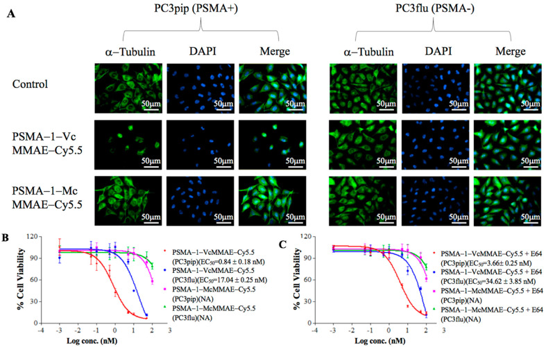
In vitro disruption of α-tubulin and cytotoxicities of PSMA-1-MMAE-Cy5.5 conjugates. (A) Immuno-detection of α-tubulin. Cells were treated with 5 nM of drugs for 24 h then fixed and stained by Alexa Fluor 488-labeled α-tubulin antibody (false color green). Selective disruption in PC3pip cells was observed by immunofluorescence only when cells were treated with PSMA-1-VcMMAE-Cy5.5. Images were taken at 40X. Representative images are shown from three independent experiments. (B) In vitro cytotoxicity of PSMA-1-VcMMAE-Cy5.5 and PSMA-1-McMMAE-Cy5.5 to PSMA-positive PC3pip cells and PSMA-negative PC3flu cells after 72-h incubation. Values are mean ± SD of six replicates. (C) In vitro cytotoxicity of PSMA-1-VcMMAE-Cy5.5 and PSMA-1-McMMAE-Cy5.5 to PC3pip and PC3flu cells after 72-h incubation in the presence of E64, a protease inhibitor. Values are mean ± SD of six replicates.

