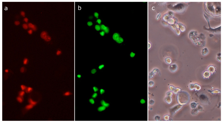
Figure 9.
Imaging of active caspases in SK-BR-3 cell line treated with 30 µM compound 4 for 6 h, using an Image-iT™ LIVE Red Poly Caspases Detection Kit: active caspases visualized using FLICA reagent (FAM-VAD-FMK poly caspases reagent) (a); dead cells permeable to SYTOX Green nucleic acid stain (b); the same area as in the previous photographs seen in phase-contrast microscopy (c).

