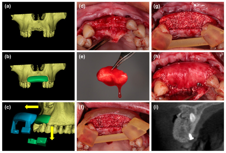
Figure 4.
(a,b) Digital simulation of bone graft contour before surgery. (c) A two-piece surgical template, which consists of two parts: the coronal part (green) for retention and the labial part (blue) for shaping the bone grafts, was fabricated based on the digital model. the template can be removed without disrupting the graft material. (d) The labial defect could be observed. (e) Particulate bone substitutes were mixed with i-PRF. (f,g) The mixture of i-PRF and particulate bone substitutes was placed into the defect under the guidance of a surgical template to form customized sticky bone. (h) The customized sticky bone was covered with a collagen membrane and fixed with pins. (i) Radiographic view of cone beam CT immediately after wound closure.

